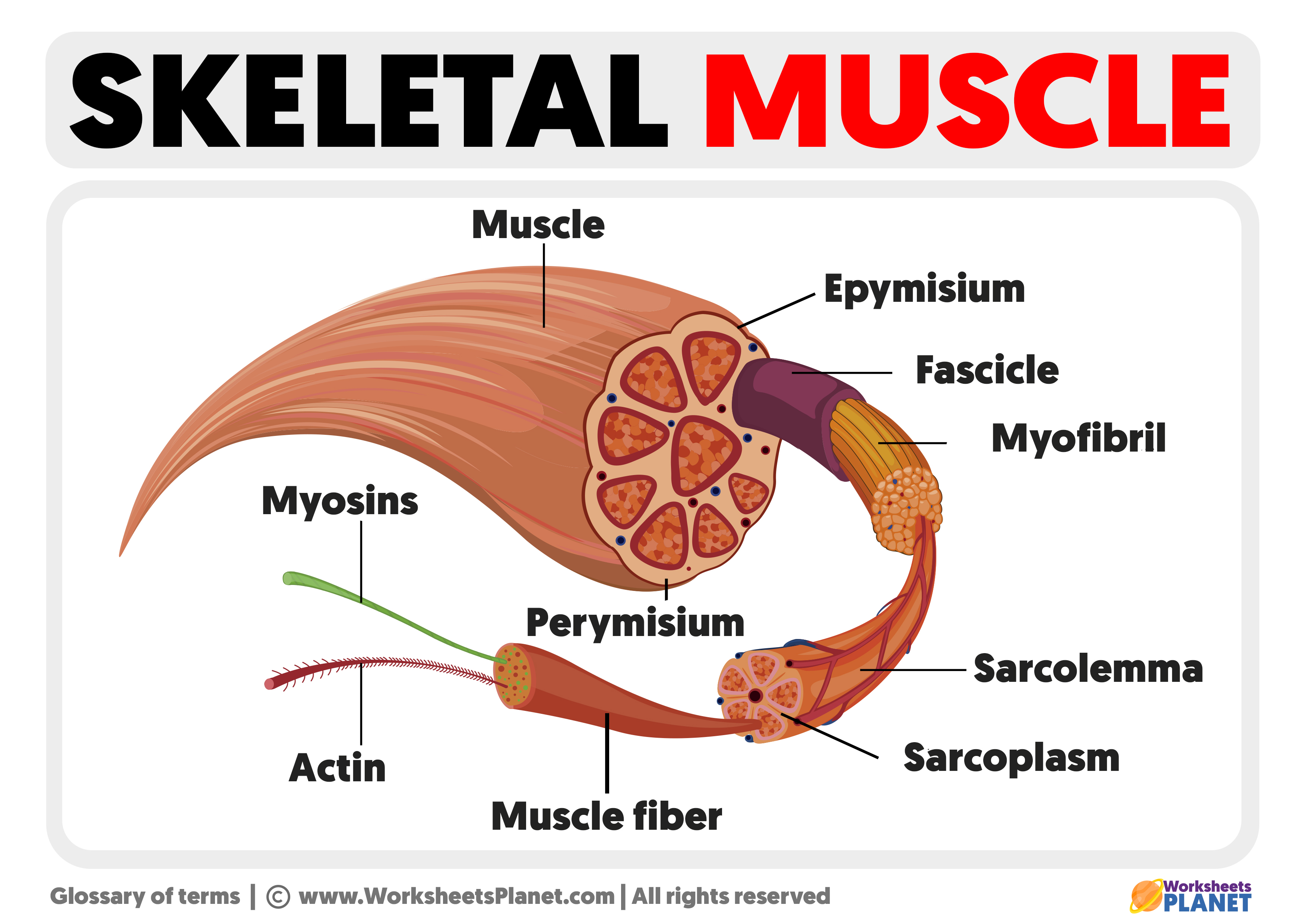
Skeletal muscle is made up of muscle fibers, surrounded by a layer of connective tissue, called the endomysium. The fibers are gathered into primary fascicles, surrounded by another layer of connective tissue, this time thicker, called the perimysium. The primary fascicles are grouped into secondary ones, protected by the epimysium, the thickest layer of connective tissue.
The epimysium continues to form the tendons and aponeurosis. Tendons and aponeuroses are made up of fibrous connective tissue. Their function is to attach muscle to bone.
The arteries, veins, and lymphatic vessels that reach the muscle must pass through the layers of connective tissue. They carry food and oxygen, necessary for muscle function. The nerves responsible for muscle activity join this structure through the motor plates, the areas where synapses are produced.

The Muscle tissue
All the muscles part of the musculoskeletal system are made up of the same types of tissues. The tissue that provides the property of contraction is the striated skeletal muscle tissue.
It is formed by fibers, resulting from the association of several cells with several nuclei, with which long structures are formed. This tissue is characterized by contracting voluntarily and quickly, since the Central Nervous System controls it.
It is called striated because of its appearance under an optical microscope. Alternate light bands are observed, called Bands I (Isotropic: they let light pass through uniformly), and dark bands, called Bands A (Anisotropic: they do not let light pass through). Thus, they constitute a repeated structure called a sarcomere, made up of proteins called actin and myosin, with the ability to contract.
The structure of the sarcomere is the one represented in the animation. Band I is made up of actin. Band A is formed by myosins and actin fragments that are introduced between them. The area where actins do not appear in Band A is observed more clearly. This band is called Band H (Hell: pale, in German).
When contraction occurs, the size of Band I and Band H decreases since the actins move closer to the center of Band A, spending chemical energy. Thus, the sarcomeres shorten, and the entire muscle shortens, producing movement.

